Contents
Scroll to:
https://doi.org/10.17073/0368-0797-2026-2-199-206
Scroll to:
Plasma surfacing in a nitrogen environment on medium-carbon steel 30KhGSA was used to form a deposited layer of high-speed molybdenum steel with a thickness of 9 – 10 mm. The authors utilized the methods of modern physical materials science to study the structural-phase states and defective substructure, mechanical and tribological properties of the surface after double high-temperature tempering and electron-beam processing with low-energy high-current beams. It is shown that the deposited layer in the initial state has a polycrystalline structure and contains eutectic interlayers. Double high-temperature tempering of the deposited layer does not change the morphology of the structure formed by eutectic grains and grains of a solid solution based on α-iron (bcc crystal lattice). The main phases are α-Fe (85 wt. %) and complex carbides Me23C6 (9 wt. %) and Me6C (6 wt. %), which form eutectic grains. High-temperature tempering of the deposited layer is accompanied by additional transformation of the residual austenite with the formation of nanosized particles of iron and chromium carbides along the boundaries of martensite crystals. After irradiation with pulsed electron beams, a structure of high-speed cellular crystallization is formed with cell sizes varying within 0.15 – 0.25 μm. Two isolated areas are distinguished, in one of which the cell boundaries do not contain precipitates of the second phase. In the cells of the second type, carbide phase interlayers are located along the boundaries - carbides of complex composition such as Me23C6 , chromium carbides Cr3C2 and molybdenum carbides MoC. Their size varies within 25 – 43 nm. The authors made a comparative analysis of mechanical and tribological properties of the surface layer of high-speed molybdenum steel after formation, tempering and electron-beam processing.
Yur’ev A.B., Gromov V.E., Baklushina I.V., Ivanov Yu.F., Kryukov R.E. Plasma coating of high-speed molybdenum steel after tempering and electron-beam processing: Structural-phase states and properties. Izvestiya. Ferrous Metallurgy. 2026;69(2):199-206. https://doi.org/10.17073/0368-0797-2026-2-199-206
Introduction
Over the past century since the development of the classical high-speed steel R18, dozens of new steels have been created for specific applications in the machine-building, metallurgical, and mining industries [1], and these have been included in handbooks and catalogs of leading foreign countries. Recently, in high-speed steels, relatively expensive and scarce tungsten has been replaced by molybdenum, which exerts a similar effect on the formation of structure and properties [2]. High-speed molybdenum steels exhibit higher performance properties than tungsten or tungsten–molybdenum steels [3 – 5].
An important challenge in improving the reliability and service life of machine and mechanism components is the purposeful and controlled modification of the properties of their working surfaces. The creation of coatings subjected to mechanical loading, wear, corrosion, and aggressive environments, while possessing high functional properties, is a fundamental task of great practical importance [6 – 8]. One approach to addressing this challenge is plasma surfacing using nitrogen as an alloying element [9 – 13].
Improvement of surface quality and its tribological and mechanical properties can be achieved by subsequent electron-beam processing. Electron-beam processing using low-energy high-current electron beams is carried out in a pulse-periodic mode, which, on the one hand, makes it possible to increase the time during which the surface layer remains molten and to homogenize its elemental composition and, on the other hand, to preserve the hardening effects leading to the formation of a submicro- and nanosized structure [14 – 17].
A fundamentally important feature of surface-layer modification by low-energy high-intensity electron beams is the absence of a pronounced interface between the modified layer and the bulk of the material. This determines the good damping properties of the material under external mechanical and thermal loading and prevents the premature initiation and propagation of brittle microcracks from the surface into the bulk of the material, which lead to fracture [18; 19].
The lack of studies clarifying the physical nature and mechanisms of structural-phase transformations in plasma-surfaced high-speed molybdenum steel coatings during deposition and subsequent treatment considerably restricts their practical application.
Materials and methods
Samples were prepared by plasma surfacing onto 30KhGSA steel in a nitrogen atmosphere using MoCrCoC flux-cored wire. The deposited layer was formed under the following conditions: welding current, 145 – 150 A; arc voltage, 50 – 55 V; surfacing speed, 18 m/h; wire feed rate, 60 m/h; and arc length, 20 mm. Surfacing was performed in four layers to a total thickness of 9 – 10 mm.
Irradiation of the deposited layer was carried out using the SOLO facility (Institute of High Current Electronics, Siberian Branch of the Russian Academy of Sciences) under the following conditions: electron energy, 18 keV; electron beam energy density, 30 J/cm2; pulse duration, 50 μs; number of pulses, 10; pulse repetition rate, 0.3 s–1; and residual gas pressure (argon) in the chamber, 0.02 Pa.
The phase composition of the deposited layer was analyzed by X-ray diffraction using a DRON-8N diffractometer. Phase identification and qualitative and quantitative phase analysis, as well as refinement of structural parameters, were performed using the KDA – Crystallography and Diffraction Analysis software package with an integrated powder diffraction database (Burevestnik JSC, version 2023-01-24-144022.8dec10c0f).
The structure and elemental composition of the deposited layer were examined by scanning electron microscopy using a KYKY-EM6900 microscope equipped with a thermionic tungsten cathode and an X-ray microanalysis attachment.
The defect substructure and the elemental and phase compositions of the deposited layer were investigated by transmission electron microscopy of thin foils in both transmission and scanning modes using a JEOL JEM-2100 microscope (Japan) [20 – 22]. Microdiffraction combined with dark-field analysis was used to determine the phase composition of the material.
The hardness and Young’s modulus of the deposited layer were measured using a NanoScan Scanning Nano-Hardness Tester at an indenter load of 0.1 N. The tribological properties of the deposited layer were evaluated in a pin-on-disc configuration under circular sliding using an Oscillating TRIBOtester tribometer (ASTM G99). Wear track profiles were recorded with a contact profilometer attached to the Oscillating TRIBOtester tribometer.
Results and discussion
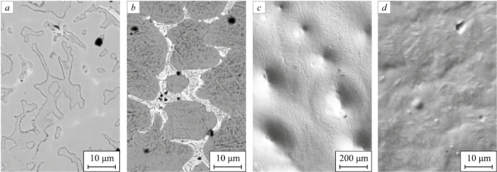
The structure of the deposited metal consists of eutectic grains and grains of a solid solution based on iron (Fig. 1, a). Double tempering of the deposited layer did not alter the polycrystalline state of the material; as in the initial state, the deposit retained a structure consisting of eutectic grains and grains of a solid solution based on iron (Fig. 1, b). Subsequent irradiation of the deposited layer with a pulsed electron beam resulted in a marked change in the morphology of the surface layer (Fig. 1, c, d). First, numerous craters formed on the deposit surface (Fig. 1, c). Second, a fine-grained structure with grain sizes on the order of several micrometers developed (Fig. 1, d). Third, the eutectic structure was no longer observed. These changes should be accompanied by modifications in the phase composition of the surface layer.
Fig. 1. Structure of the surface layer of molybdenum high-speed steel deposited |
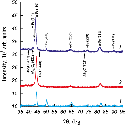
This assumption is supported by the results of X-ray diffraction analysis, which revealed a significant change in the phase composition of the deposited material after irradiation with pulsed electron beams (Fig. 2; Table).
Fig. 2. Fragments of radiographs of the plasma coating in the initial state (1),
Calculation results obtained from diffraction patterns
| |||||||||||||||||||||||||||||||||||||||||||||||||||||||||||||||
Comparison of the data presented in the Table shows that both tempering of the deposited material and subsequent irradiation of the tempered material with pulsed electron beams led to a substantial decrease in the carbide-phase content, from 23 wt. % in the initial deposit to 3 wt. % after irradiation. Tempering of the deposited layer was accompanied by additional transformation of the residual austenite, whereas subsequent irradiation again led to the retention of residual austenite in the surface layer. It may be assumed that the decrease in the relative carbide-phase content after tempering, and especially after additional irradiation, was associated with the formation of a supersaturated solid solution based on α- and γ-iron. However, the crystal lattice parameter of α-iron was highest in the initial state and lowest after additional irradiation (see Table). This finding indicates, among other things, the release of alloying elements from the crystal lattice of α-iron, which in turn suggests the formation of carbide-phase particles. These apparently contradictory results may indicate that the second-phase particles formed during additional treatment of the deposited metal are nanosized.
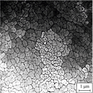
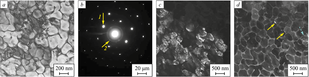
STEM analysis showed that the structure of the surface layer after irradiation with a pulsed electron beam consisted of high-speed crystallization cells ranging in size from 0.15 to 0.25 μm (Fig. 3).
Fig. 3. Surface structure of the deposited layer irradiated |
Two isolated areas containing different types of high-speed crystallization cells were identified. In the first type of cells, the boundaries were nearly free of second-phase precipitates, whereas in the second type, extended interlayers of the second phase were located along the boundaries. The sizes of these inclusions ranged from 25 to 43 nm. It may be assumed that the first-type crystallization cells formed as a result of high-speed crystallization in molten regions corresponding to grains of a solid solution based on α-iron, whereas the second-type crystallization cells formed during crystallization of molten eutectic grains. This explains the marked difference in the content of second-phase particles between the two cell types.
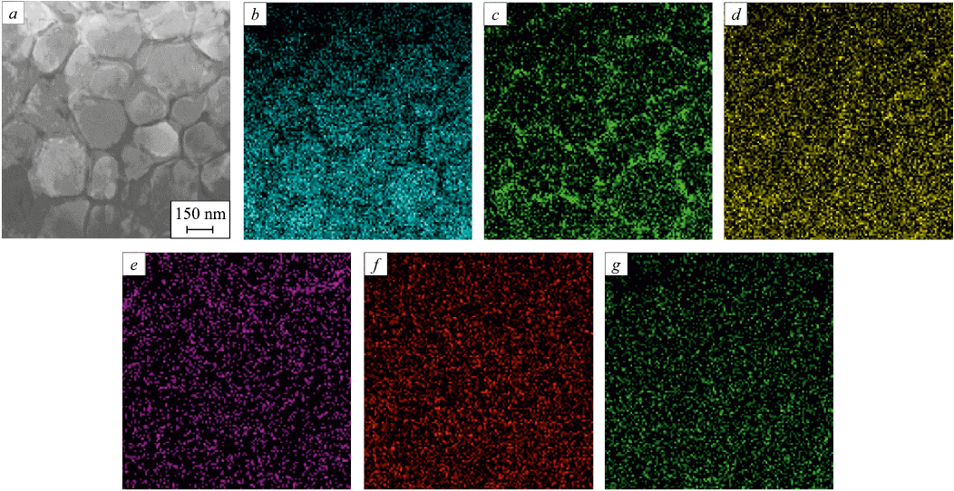
X-ray microanalysis showed that the interiors of crystallization cells of both types were enriched predominantly in iron atoms (Fig. 4, b), whereas extended interlayers enriched predominantly in molybdenum (Fig. 4, c), chromium (Fig. 4, d), and carbon (Fig. 4, e) were detected along the cell boundaries.
Fig. 4. TEM image of a section of the deposited layer surface (a) and images of this section obtained |
Dark-field analysis and indexing of TEM electron diffraction patterns were used to determine the phase composition of the high-speed crystallization cells. In the first-type crystallization cells, the cell interior was found to consist of a solid solution based on α-iron. In some cases, nanosized grains of residual austenite as well as nanosized particles of carbides of the Me23C6 and MoC types were detected (Fig. 5). A dislocation substructure in the form of randomly distributed dislocations was observed within the crystallization cells.
Fig. 5. TEM image of the structure of the first type of cells formed after irradiation |
Analysis of the phase composition of surface-layer regions containing second-type crystallization cells showed that the cell interior also consisted of a solid solution based on α-iron. In some cases, reflections corresponding to residual austenite were detected in the electron diffraction patterns. Numerous second-phase inclusions were located along the crystallization cell boundaries, and analysis of the corresponding electron diffraction patterns indicated that they were carbides of the Me23C6 , Cr3C2 and Cr7C3 compositions.
Following this combined treatment of the surface layer of the high-speed molybdenum steel, both the mechanical and tribological properties changed. Additional irradiation of the deposited layer with a pulsed electron beam reduced the wear parameter and the coefficient of friction from k = 2.0·10–6 mm3/(N·m) and μ = 0.75 for the deposit in the initial state to k = 1.25·10–6 mm3/(N·m) and μ = 0.60 for the deposit after tempering and additional pulsed-electron-beam irradiation. For the substrate (30KhGSA steel), k = 10.0·10–6 mm³/(N·m) and μ = 0.44. Thus, irradiation of the deposited layer with pulsed electron beams resulted in a slight decrease in the wear parameter by a factor of 1.6 and in the coefficient of friction by a factor of 1.23. Compared with 30KhGSA steel, the wear parameter decreased eightfold, whereas the coefficient of friction increased by a factor of 1.36.
The nanohardness of the surface layer of the deposited high-speed molybdenum steel after irradiation with pulsed electron beams ranged from 9.5 to 14.7 GPa and averaged 10.7 GPa within a surface layer 100 μm thick. The mean nanohardness of the deposited layer in the initial state was 12.4 GPa. Thus, irradiation of the deposited high-speed molybdenum steel with pulsed electron beams resulted in a 1.16-fold decrease in nanohardness. The nanohardness of the substrate (30KhGSA steel) was 3.2 GPa and was substantially lower, by a factor of 3.3, than the mean hardness of the deposited metal after irradiation with a pulsed electron beam.
The Young’s modulus of the surface layer of the high-speed molybdenum steel irradiated with pulsed electron beams, averaged over 10 indents, was 215 GPa. Given that the mean Young’s modulus of the deposited metal in the initial state was 234.9 GPa, irradiation of M9 high-speed molybdenum steel resulted in a slight decrease in this parameter by a factor of 1.09.
Conclusions
A 9 – 10 mm thick deposited layer of high-speed molybdenum steel was formed on medium-carbon 30KhGSA steel by plasma surfacing in a nitrogen alloying atmosphere. The deposited layer was then subjected to double tempering followed by irradiation with pulsed electron beams. Mechanical and tribological tests were carried out for the deposited layer in the initial state, after tempering, and after electron-beam processing. After irradiation, the nanohardness of the deposited layer was found to be 3.3 times higher than that of 30KhGSA steel. The wear parameter of the irradiated deposited layer was eight times lower than that of 30KhGSA steel, whereas its coefficient of friction was 1.36 times higher. After electron-beam processing, the high-speed crystallization cells measured 0.15 – 0.25 μm in size. Two isolated areas containing different cell types were identified. In the first-type cells, the boundaries were almost free of second-phase precipitates. In the second-type cells, extended carbide-phase interlayers were present along the boundaries. These interlayers consisted of complex carbides of the Me23C6 type, chromium carbides Cr7C3 and Cr3C2 , and molybdenum carbides MoC, with particle sizes ranging from 25 to 43 nm. The pronounced difference in the content of second-phase particles between the two cell types is attributed to their different origins. The first-type crystallization cells formed during high-speed crystallization of molten regions corresponding to grains of a solid solution based on α-iron, whereas the second-type cells formed during crystallization of molten eutectic grains.
1. Kremnev L.S. From high-speed tungsten steel to high-temperature molybdenum steel: A century of high-speed steel. Steel in Translation. 2009;39(12):1111–1118. https://doi.org/10.3103/S0967091209120195
2. Kremnev L.S., Onegina A.K., Vinogradova L.A. Special features of transformations, structure, and properties of molybdenum high-speed steels. Metallovedenie i termicheskaya obrabotka metallov. 2009;(12(654)):13–19. (In Russ.).
3. Kremnev L.S. Alloying theory and development of heat-resistant tool steels and alloys. Metallovedenie i termicheskaya obrabotka metallov. 2008;(11(641)):18–28. (In Russ.).
4. Kupalova I.K. Phase analysis and phase composition of high-speed steels (review). Zavodskaya laboratoriya. 1983; (1):27–40. (In Russ.).
5. Vostretsov G.N. Deformation ability of deposited heat-resistant metal of P2M8 type during martensitic transformation. Bulletin of the Siberian State Industrial University. 2012;(1):7–10. (In Russ.).
6. Chaus A.S., Pokorný P., Čaplovič Ľ., Sitkevich M.V., Peterka J. Complex fine-scale diffusion coating formed at low temperature on high-speed steel substrate. Applied Surface Science. 2018;437:257–270. https://doi.org/10.1016/j.apsusc.2017.12.173
7. Hashemi N., Mertens A., Montrieux H.-M., Tchuindjang J.T., Dedry O., Carrus R., Lecomte-Beckers J. Oxidative wear behavior of laser clad high speed steel thick deposits: Influence of sliding speed, carbide type and morphology. Surface and Coatings Technology. 2017;315:519–529. https://doi.org/10.1016/j.surfcoat.2017.02.071
8. Darmawan W., Quesada J., Marchal R. Characteristics of laser melted AISI-T1 high speed steel and its wear resistance. Surface Engineering. 2007;23(2):112–119. https://doi.org/10.1179/174329407X169502
9. Emelyushin A.N., Petrochenko E.V., Nefed′ev S.P. Investigation of the structure and impact-abrasive resistance of coatings of the Fe-C-Cr-Mn-Si system, additionally alloyed with nitrogen. Welding International. 2013;27(2):150–153. https://doi.org/10.1080/09507116.2012.695548
10. Nefedyev S.P., Emelyushin A.N. The influence of nitrogen on the formation of the structure and properties of plasma coatings of the 10P6M5 type. Bulletin of Ugra State University. 2021;(3(62)):33–45. (In Russ.).
11. Nefedyev S.P., Vdovin K.N., Emelyushin A.N. Peculiarities of forming of the wear-resistant cast iron coating structure on steel 45 upon plasma-powder surfacing. Materials Science Forum. 2016;870:141–148. https://doi.org/10.4028/www.scientific.net/MSF.870.141
12. Emelyushin A.N., Petrochenko E.V., Nefedyev S.P. Investigation of the structure and impact-abrasive wear resistance of Fe-C-Cr-Mn-Si system coatings additionally alloyed with nitrogen. Welding Production. 2011;(10):18–22. (In Russ.).
13. Mozgovoy I.V., Shneider E.A. Surfacing of High-Speed Steel. Omsk: OmSTU; 2016:200. (In Russ.).
14. Rotshtein V.P., Proskurovskii D.I., Ozur G.E., Ivanov Yu.F. Modification of Metallic Materials Surface by Low-Energy High-Current Electron Beams. Novosibirsk: SB RAS Nauka; 2019:348. (In Russ.).
15. Evolution of Surface Layer Structure of Steel Subjected to Electron-Ion-Plasma Processing. Koval’ N.N., Ivanov Yu.F. eds. Tomsk: NTL; 2016:298. (In Russ.).
16. Surface Modification and Alloying by Laser, Ion, and Electron Beams. Poate J.M., Foti G., Jacobson D.C. eds. NY: Springer New York; 1983:414. https://doi.org/10.1007/978-1-4613-3733-1
17. Ivanov Yu.F., Gromov V.E., Minenko S.S., Guseva T.P., Chapaykin A.S., Semin A.P. Structure and properties of the surface layer obtained by plasma surfacing from high-entropy molybdenum high-speed steel after complex processing. Materialovedenie. 2025;(6):15–23. (In Russ.). https://doi.org/10.31044/1684-579X-2025-0-6-15-23
18. Gromov V.E., Ivanov Yu.F., Yuriev A.B., Minenkо S.S., Konovalov S.V. Modification of transition zone structure of high-speed steel surfacing ‒ substrate by electron-beam treatment. Bulletin of the Siberian State Industrial University. 2025;(1):43–50. https://doi.org/10.57070/2304-4497-2025-1(51)-43-50
19. Shulov V.A., Paikin A.G., Belov A.B., L’vov A.F., Engel’ko V.I., Ovchinnikov D.V. Surface modification of the parts made from heat resistant steel by high-current pulsed electron beam. Fizika i Khimiya Obrabotki Metallov. 2005;(2):61−70. (In Russ.).
20. Egerton F.R. Physical Principles of Electron Microscopy. Basel: Springer International Publishing; 2016:196.
21. Kumar C.S.S.R. Transmission Electron Microscopy. Characterization of Nanomaterials. New York: Springer; 2014:717.
22. Carter C.B., Williams D.B. Transmission Electron Microscopy. Berlin: Springer International Publishing; 2016:518.
Aleksei B. Yur’ev, Dr. Sc. (Eng.), Prof., Rector
2/3 Akademicheskii Ave., Tomsk 634055, Russian Federation
Viktor E. Gromov, Dr. Sci. (Phys.-Math.), Prof., Head of the Chair of Science named after V.M. Finkel’
2/3 Akademicheskii Ave., Tomsk 634055, Russian Federation
Irina V. Baklushina, Deputy Director of the Institute of Open Education
2/3 Akademicheskii Ave., Tomsk 634055, Russian Federation
Yurii F. Ivanov, Dr. Sci. (Phys.-Math.), Prof., Chief Researcher
2/3 Akademicheskii Ave., Tomsk 634055, Russian Federation
Roman E. Kryukov, Dr. Sci. (Eng.), Prof. of the Chair of Mechanics and Machinery
2/3 Akademicheskii Ave., Tomsk 634055, Russian Federation
Yur’ev A.B., Gromov V.E., Baklushina I.V., Ivanov Yu.F., Kryukov R.E. Plasma coating of high-speed molybdenum steel after tempering and electron-beam processing: Structural-phase states and properties. Izvestiya. Ferrous Metallurgy. 2026;69(2):199-206. https://doi.org/10.17073/0368-0797-2026-2-199-206
 Email this article (Login required)
Email the author (Login required)
Email this article (Login required)
Email the author (Login required) Publishing House of National University of Science and Technology "MISIS"
4, Leninskii prospekt, Moscow, 119991, Russia
Phone: +7 (495) 638-44-11, +7 (495) 784-58-66
E-mail: fermet.misis@mail.ru